Summary
Interstitial lung disease (ILD) is a heterogeneous group of disorders characterized by inflammation and/or fibrosis of the pulmonary interstitium [1]. Impaired gas exchange leads to worsening respiratory symptoms and declining lung function [1]. ILD can be classified according to etiology (e.g., idiopathic, autoimmune-related, exposure-related) [2]. Usual interstitial pneumonia (UIP), nonspecific interstitial pneumonia (NSIP), and organizing pneumonia (OP) are the most frequently encountered radiological-histopathological patterns [2]. ILD commonly complicates connective tissue disease (CTD), although its prevalence and predominant pattern vary across CTDs [3]. In pooled data, UIP predominates in rheumatoid arthritis, whereas NSIP predominates in mixed CTD, systemic sclerosis, idiopathic inflammatory myositis, and primary Sjögren syndrome [3]. In patients with UIP, CTD-associated disease should be differentiated from idiopathic pulmonary fibrosis (IPF) because the distinction affects prognosis and treatment [4]. On CT, the straight-edge, exuberant honeycombing, and anterior upper lobe signs are suggestive of CTD-associated ILD (CTD-ILD) [5].
Definition and classification
ILD has been defined as “a heterogeneous and complex group of conditions characterized by inflammation, fibrosis, or both, in the interstitium of the lungs” [1].
ILD can be classified by etiology into idiopathic interstitial pneumonia, autoimmune-related ILD, exposure-related ILD, ILD with cysts or airspace filling, or ILD related to distinct primary diseases (e.g., sarcoidosis) [2]. A residual group includes acute and chronic eosinophilic pneumonia and malignant disease–associated ILD [2].
Radiological-histopathological patterns
UIP, NSIP, and OP are the most frequently encountered patterns [2].
Usual interstitial pneumonia (UIP)
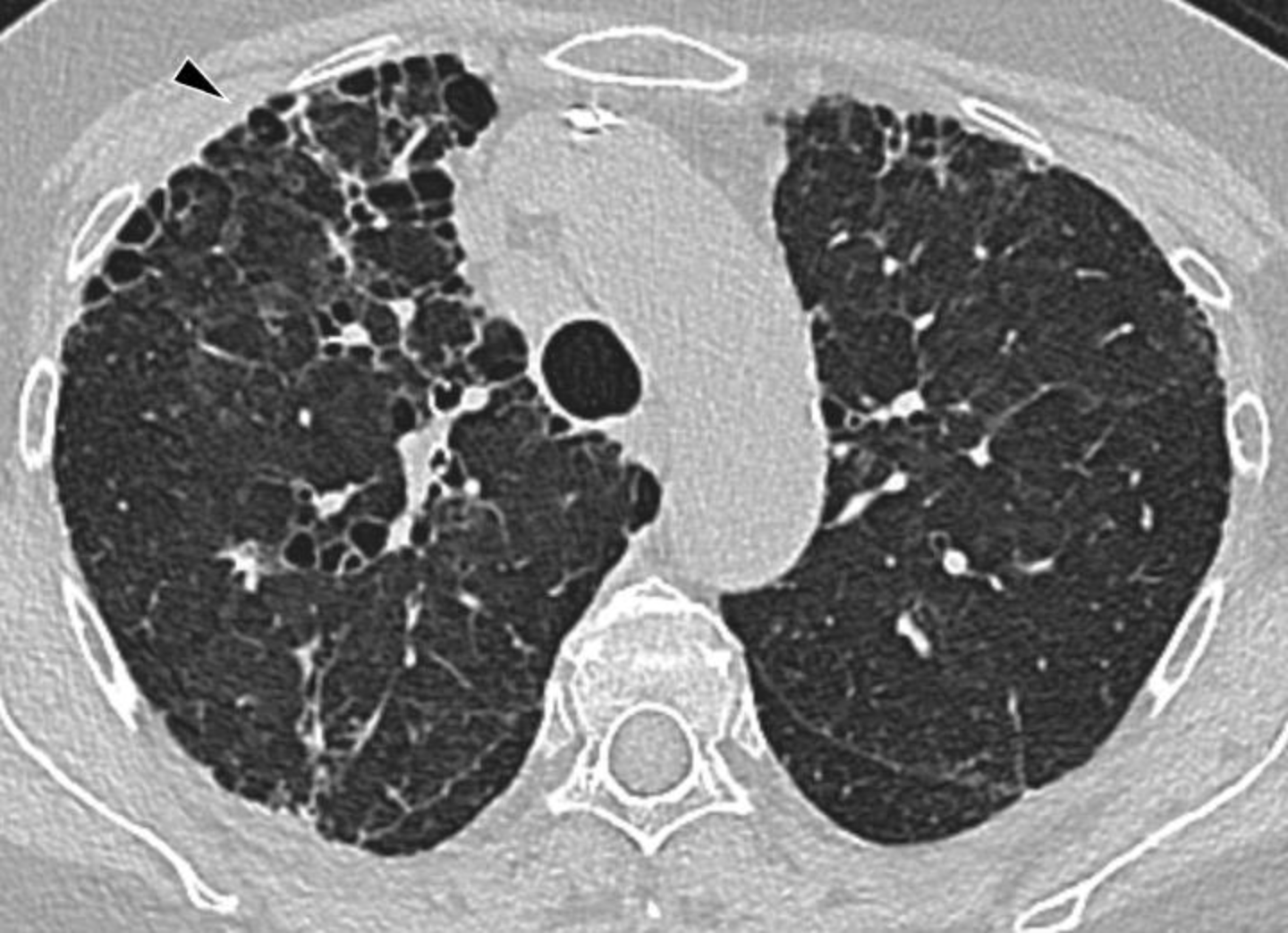
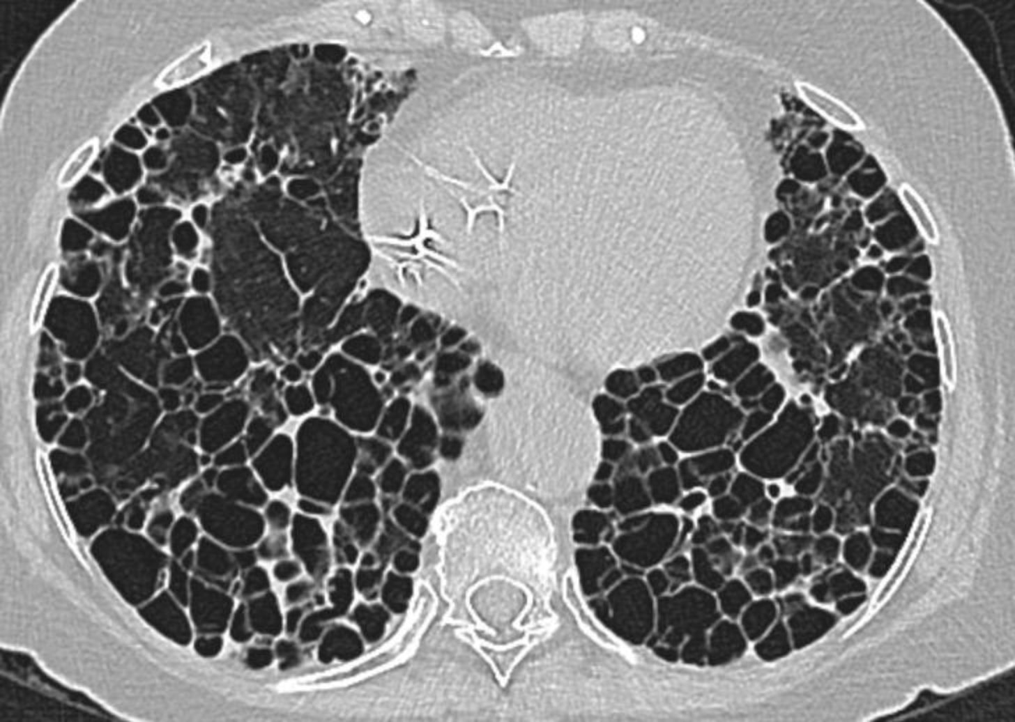
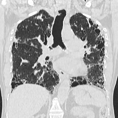
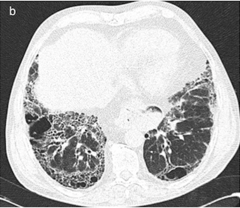
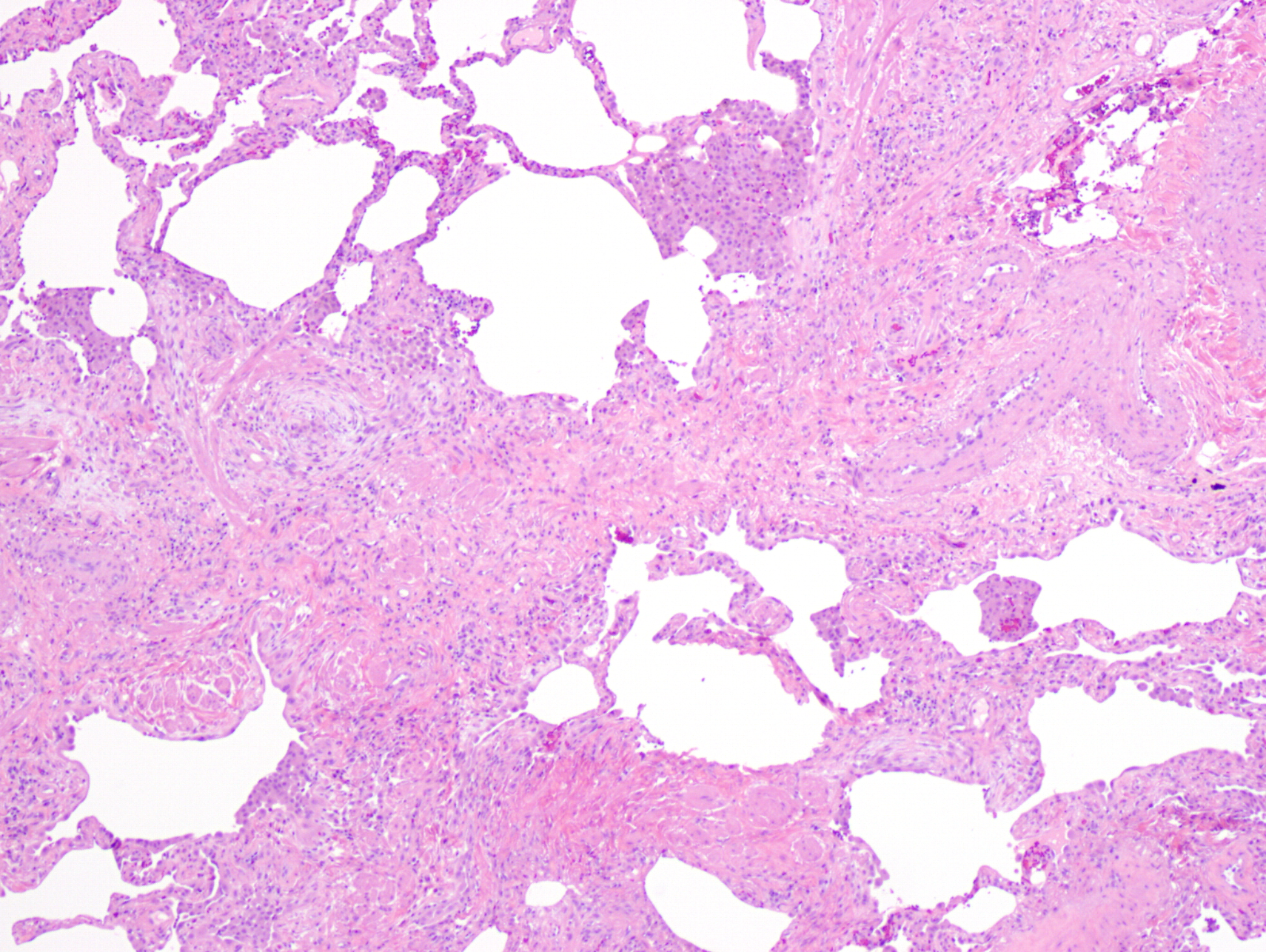
On CT, UIP is characterized by reticular opacities and honeycombing with basal and subpleural predominance and architectural distortion [6]. Histologically, UIP shows patchy, dense fibrosis that distorts the lung architecture (“temporal heterogeneity”) [7].
Nonspecific interstitial pneumonia (NSIP)
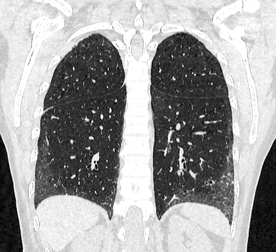
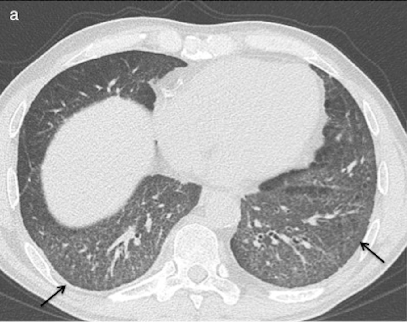
On CT, NSIP shows reticular and ground-glass opacities with basal and subpleural predominance [8]. In contrast to UIP, the lung architecture is preserved [8]. Immediate subpleural sparing “is present in a minority (~30%), but when present, is a key finding of NSIP” [8]. Histologically, NSIP shows diffuse, uniform inflammation (“temporal homogeneity”) [9].
Organizing pneumonia (OP)
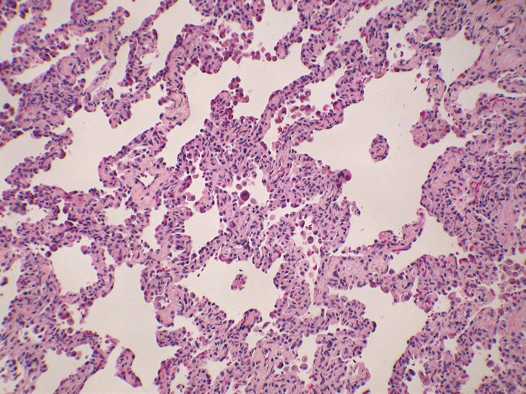
On CT, OP appears as ground-glass opacity with air bronchograms in a bronchocentric distribution [10]. In one series, the reversed halo sign was present in 6 of 31 patients (19%) with biopsy-proven cryptogenic organizing pneumonia (COP) [11]. This frequency may not be representative, because only biopsy-proven cases were included [11]. Histologically, OP is characterized by fibroblastic plugs within alveolar sacs, ducts, and bronchiolar lumen [12].
Connective tissue disease–associated ILD
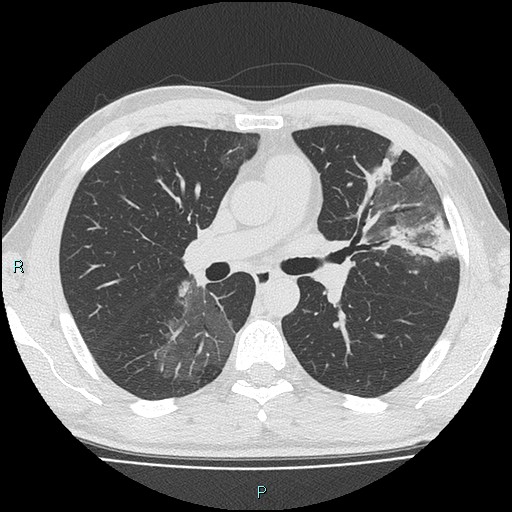
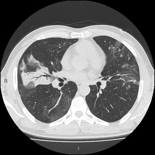
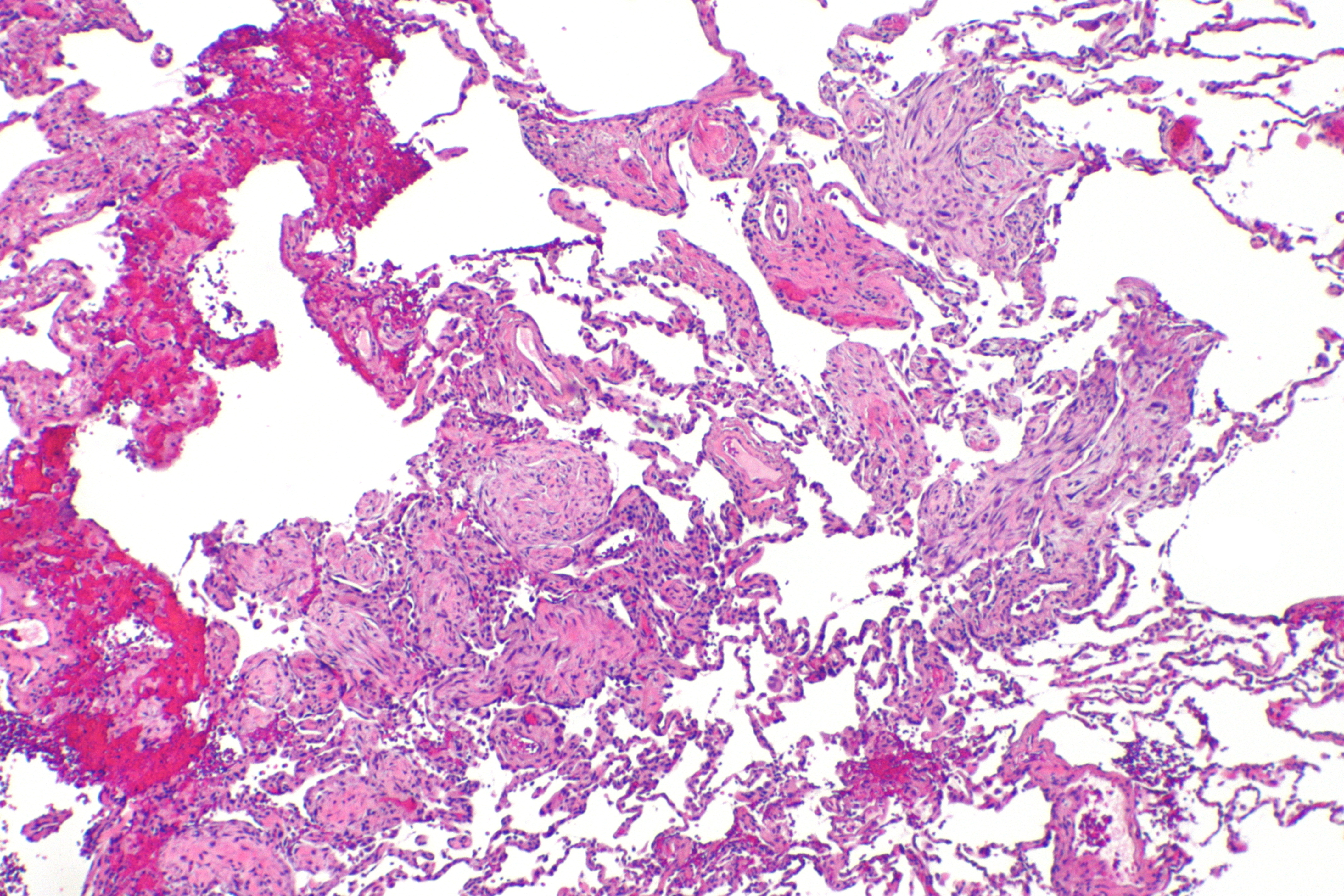
ILD commonly complicates CTD and is reported in every CTD, although its prevalence and pattern vary considerably across CTDs [3]. Yoo et al. [13] note that “ILD exists in approximately 40–50% of patients with CTDs, and is the main cause of morbidity and mortality.”
Deba Ayarza et al. [4] stress the clinical consequence: “In patients with UIP, it is important to differentiate those secondary to CTD from those with IPF. This distinction is of significant prognostic and therapeutic value, as secondary pulmonary involvement may benefit from treatment of the systemic disease to attempt to prevent progression.”
Pooled prevalence of ILD according to CTD
Joy et al. [3] reported the pooled prevalence of ILD and the most common pattern for each CTD.
| CTD | Pooled prevalence of ILD | Most common pattern |
|---|---|---|
| Mixed connective tissue disease | 56% | NSIP |
| Systemic sclerosis | 47% | NSIP |
| Idiopathic inflammatory myositis | 41% | NSIP |
| Primary Sjögren syndrome | 17% | NSIP |
| Rheumatoid arthritis | 11% | UIP |
| Systemic lupus erythematosus | 6% | Other/undifferentiated |
CT signs suggestive of CTD-ILD
In patients with a UIP pattern, three CT signs are suggestive of CTD-ILD [5].
| Sign | Definition | Specificity | Sensitivity |
|---|---|---|---|
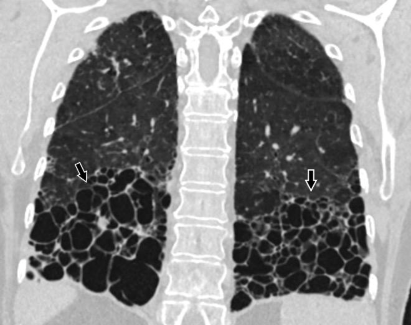
| Straight-edge | Fibrosis confined to the lung bases that ends abruptly in the craniocaudal direction, with no substantial spread up the lateral lung margins on coronal images | 94.0% | 25.4% |
| Exuberant honeycombing | Honeycomb cysts making up > 70% of the fibrotic lung | 94.0% | 22.2% |
| Anterior upper lobe | Upper lobe fibrosis concentrated anteriorly, with the rest of the upper lobes relatively spared, together with lower lobe involvement | 87.2% | 25.4% |
In a published case with a UIP pattern, all three signs were present [4].